Optical Projection Tomography as a Novel Method to Visualize and Quantitate Whole-Brain Patterns of Cell Proliferation in the Adult Zebrafish Brain
Optical Projection Tomography as a Novel Method to Visualize and Quantitate Whole-Brain Patterns of Cell Proliferation in the Adult Zebrafish Brain
Benjamin W Lindsey and Jan Kaslin
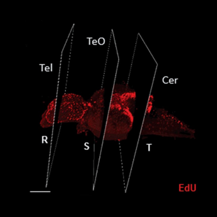
The zebrafish has become a popular model in neuroscience to study changes arising in the mature central nervous system (CNS) as a result of social behaviors, neurogenesis, brain injury and disorders, regeneration, neuroimmune interactions, and neurodegenerative diseases. Unfortunately, few brain-wide imaging tools are available to researchers investigating changes in the CNS during adulthood. Most studies continue to be performed by the standard method of fixing whole adult brains, sectioning, and confocal imaging antibody markers of interest. However, serial sectioning is time-consuming and severely limits interpretations of changes occurring across the entire brain axis. By contrast, the ability to visualize markers in threedimensional (3D) space allows investigators to screen the adult brain for regions of interest following manipulation. In this study, we have developed an efficient pipeline to clear, visualize, and quantitate the whole adult zebrafish brain using optical projection tomography (OPT) to assess changes in cell proliferation alone or in combination with transgenic reporter lines.
READ FULL PUBLICATION
Lindsey and Kaslin, 2017 (413 KB)


